

You deserve more.
Put the Power of Artificial Intelligence Behind Your Mammogram
Lenox Hill Radiology Centers
Lenox Hill operates convenient outpatient imaging centers located in the metropolitan New York area. Our goal is to provide state of the art imaging services coupled with accurate diagnosis and efficient services, including MRI, CT, Digital & 3D Mammography, Ultrasound, PET-MRI, PET-CT, Nuclear Medicine, Fluoroscopy, X-ray, DEXA | Bone Density, and more.
Our combination of dedicated specialty physicians, friendly compassionate staff and cutting edge technology allow us to offer our patients only the very best in diagnosis, management, follow up and care.


Trending Radiology
Recent Radiology News
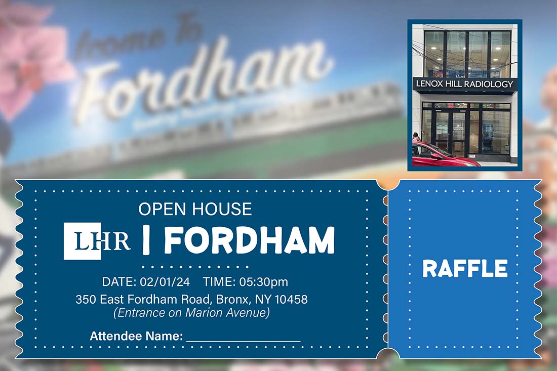

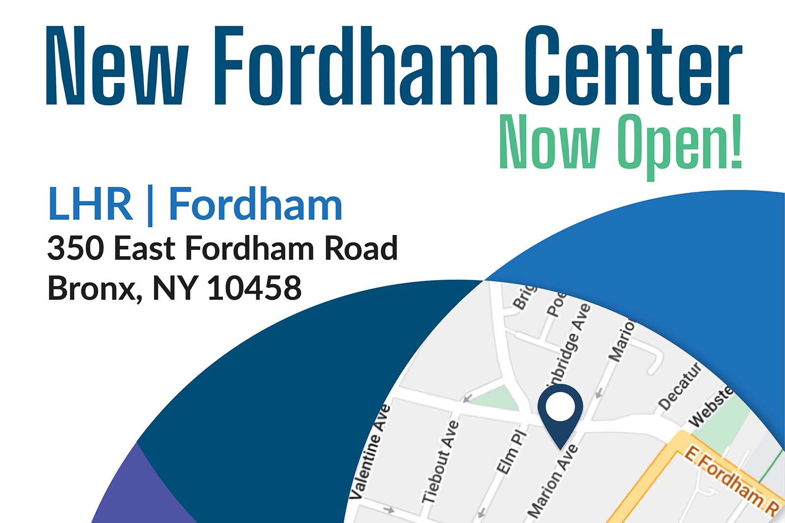
Imaging Centers Near You
See what services are performed at each of our locations.
Radiology Centers Near You

Flexible hours designed around your schedule.
Appointments available on your terms.

